Decision making and techniques to simplify dental extractions in pet dogs have actually been described.1-5 Proper perioperative planning and decision making regarding canine extractions can improve surgical result.
Preoperative Factors To Consider in the Canine Dental Client
It is necessary to properly assess the canine patient prior to the performance of extractions. This consists of total general physical and oral assessments and suitable preoperative blood work. Once the client has actually been appropriately assessed it is essential to pick a suitable anesthetic protocol that will offer the canine oral client with sufficient perioperative discomfort management.
Oral Evaluation in the Awake Dog Patient
Oral assessment in the awake canine client resembles the oral assessment in the awake feline patient. Abnormalities detected are talked about with the owner with the specification that additional abnormalities might be found in the anesthetized client.
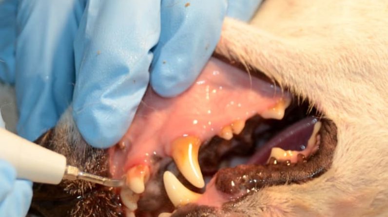
Oral Examination in the Anesthetized Dog Client
Oral assessment in the anesthetized canine client begins with a comprehensive oral examination consisting of assessment for missing or supernumerary teeth, malformed teeth, proper occlusion, periodontal probing and expedition of the teeth with an oral explorer to spot pulpal exposure, worn teeth and dental caries. Abnormalities are noted on the canine oral chart.
Dental Radiography in the Dog Patient Prior to Extraction
Oral radiography is an essential tool in the decision making process in canine dental clients. Dental radiography can help identify the most appropriate treatment technique in canine teeth impacted with periodontal disease, endodontic disease, dental caries and other sores.
A dental radiograph taken prior to carrying out a hard extraction will supply the veterinarian with crucial information regarding the tooth. Radiographic assessment of the tooth will figure out if other treatment alternatives might be possible so that the owner can be used options to extraction. In cases of extreme periodontal or endodontic illness extraction might be the best treatment alternative for the patient. Oral radiographs prior to extraction will also reveal structural abnormalities that might be present in the tooth or surrounding bone. These structural irregularities consist of: severe periradicular bone loss secondary to gum or endodontic illness, supernumerary roots, unusual root angulation including convergent roots and excessive curvature of the apical portion of the root, ankylosis and hypercementosis. Understanding of these structural abnormalities prior to initiation of the extraction will provide essential info regarding the most suitable strategy for the extraction and will help in reducing the occurrence of problems.
Appropriate Equipment and Instrumentation for Canine Extractions
A high-speed handpiece with fiberoptics is very practical when performing surgical extractions in pet dogs. The fiberoptic handpiece provides a light directly on the surgical website. Burs used regularly consist of a variety of round burs for the elimination of buccal bone and tapered crack burs for sectioning multi-rooted teeth. Necessary hand instrumentation for performing canine extractions have actually been formerly described.1-5 Hand instrumentation particularly created for canine extractions is offered through various veterinary supply business. Instruments for canine extractions might be packaged together in a canine extraction pack and steam sanitized prior to each usage. Instrumentation in canine extraction packs consist of: scalpel manage upon which a # 15 blade can be positioned prior to surgical treatment, a periosteal elevator, a soft tissue retractor, a variety of oral elevators and luxators, extraction forceps, needle holders, Adson tissue forceps, suture scissors and an iris scissors for cutting soft tissue. A little root forceps is also valuable for reaching down into an alveolus and obtaining a firm grasp on a loose root idea. It is important to consistently sharpen dental extraction instrumentation to guarantee ideal performance.
Structural Features of Canine Teeth
The oral formula in the adult dog is: 2 (I 3/3, C1/1, P4/4, M2/3) = 42. The incisors and canine teeth all have one root. The 1st premolars and the lower 3rd molars have one root. The upper 2nd and 3rd premolars and the lower second,3 rd fourth premolars and 1st and second premolars have 2 roots and the upper 4th premolar and 1st and second molars have 3 roots. Knowledge of the location of the furcation of the teeth will allow precise sectioning of teeth during surgical extractions.
Techniques for Performing Simple and Multi-Rooted Extractions in Canines
There are numerous different techniques for performing extractions in the pet. These methods consist of an easy extraction, multi-rooted extraction and surgical extraction
Simple or Closed Extraction
The incisors, the maxillary and mandibular 1st premolars and the mandibular 3rd molar are typically small single rooted teeth in the canine and can be normally be removed using basic or closed extraction techniques. Easy or closed extraction strategies have actually been previously described.1-5 The procedure is started by cutting the gingival attachment around the entire area of the tooth using a No. 11 scalpel blade in a handle or a sharp luxator. A luxator that matches the curvature of the tooth is picked and is put into the gingival sulcus at a small angle to the tooth and pressed into the periodontal ligament space and worked around the whole circumference of the tooth using mild apical pressure. The operator might now choose to continue the extraction utilizing an oral elevator or continue utilizing a luxator. A dental elevator may be utilized when sufficient area has actually been developed for the thicker tipped instrument. A suitable sized elevator is chosen, placed in the gum ligament area and worked around the tooth with a gentle rotational pressure held at each point for 10-15 2nd to assist break down the periodontal ligament. When the tooth ends up being loose it can be gotten rid of digitally or gently understood with a dental extraction forceps placed as far apically on the tooth as possible and with a gentle rotational movement of the forceps in the long axis of the tooth, the tooth may be turned and gotten rid of from the alveolus.
Multi-Rooted Extraction
Extraction of multi-rooted teeth in dogs starts by cutting the gingival attachment to the tooth with either a No. 11 or 15 scalpel blade in a deal with or a properly sized sharp luxator. The furcation(s) of the tooth lie utilizing visual examination of the gingiva and alveolar crest. Furcations might be found by observing where the gingiva and alveolar crestal bone raises a little coronally. Elimination of a small amount of bone in this location with a round bur will help imagined the furcation. Once the furcation is pictured the tooth is sectioned by positioning a tapered fissure bur (# 701 or # 701L) at the furcation and sectioning the tooth through the crown. One cut is made in 2 rooted teeth and two cuts are made in 3 rooted teeth to divide the tooth into several single systems. To validate that the tooth has actually been successfully sectioned, a dental elevator is positioned between the sectors and carefully rotated. If the sections move somewhat apart then the sectioning is total; if the sectors do stagnate following minor take advantage of between the cusp segments then the sectioning is most likely to be incomplete and addition burring is necessary to finish the sectioning. Once the sectioning is total the specific roots are extracted individually as formerly explained for simple extractions.
Techniques for Performing Surgical Extractions in Pet Dogs
A complex or surgical extraction technique is usually booked for teeth that are difficult to extract since of their big root structure consisting of the canine teeth, mandibular first molars and the maxillary 4th premolars. A surgical extraction might likewise be carried out when teeth are ankylosed or when trying to retrieve a broken root idea. The teeth most typically needing surgical extractions consist of the canine teeth and the carnassial teeth.
Surgical Extraction of Canine Teeth
Surgical extraction of the maxillary canine tooth is initiated by making divergent incisions mesial and distal to the canine tooth and producing a mucoperiosteal flap. The buccal alveolar bone is removed as needed with a big round bur to quickly extract the tooth with luxators and dental elevators. Care must be taken to prevent producing an oronasal fistula during the extraction. The periosteal layer of the flap is incised apically to ease stress on the flap prior to closure.
There are two methods for the surgical extraction of the mandibular canine teeth consisting of the labial and linguistic technique. The labial technique utilizes a mucoperiosteal flap situated on the labial element of the tooth while a linguistic approach uses a lingually located flap. Equal quantities of alveolar bone are present buccally and labially so there is no advantage of one strategy over the other with regard to bone elimination. The psychological artery, vein and nerve exit through the mental foramen located near the labial aspect of the apex of this tooth. A linguistic approach prevents prospective damage to these structures.
Surgical Extraction of the Maxillary 4th Premolars
When carrying out a mucoperiosteal flap for the surgical extraction of the maxillary 4th premolar a number of structures ought to be thoroughly prevented. When making the mesial (rostral) portion of the incision the infraorbital artery, vein and nerve need to be avoided as they exit the infraorbital canal right away rostral to the periapical bone of the mesiobuccal root of the maxillary fourth premolar. These structures can be avoided by digitally retracting them dorsally and not extending this cut too far apically. When making the distal (caudal) part of the cut the parotid and zygomatic salivary duct papillae should be pictured and avoided. After raising the mucoperiosteal flap the furcations lie utilizing a round bur. The tooth is then sectioned through the furcation in between the mesiobuccal and distal roots with a # 701L tapered crack bur from the furcation through the crown. Alveolar bone over the distal root is eliminated as required to remove the distal root. At this moment some operators choose to cut off part of the staying portion of the crown. The bur is put in the furcation perpendicular to the tooth at the base of the palatal wall of the mesiobuccal cusp to section the mesiobuccal and palatal roots. The alveolar bone over the mesiobuccal roots is gotten rid of as required to get rid of the mesiobuccal root. The interradicular bone between the mesiobuccal and palatal roots can be eliminated as required to expose the palatal root. When extracting the palatal root it is essential to direct the luxator in a somewhat palatal direction to follow the palatal instructions of the peak of this root. The extraction website is débrided, flushed and closed in a regular way.
Surgical Extraction of the Mandibular 1st Molars
The surgical extraction of the mandibular 1st molar is initiated with a mucoperiosteal flap with two divergent releasing cuts on the mesial and distal element of the buccal aspect of the tooth. The mucoperiosteal flap is raised and the furcation lies and sectioned. The distal and mesial edges of the cusps of the tooth may be removed to provide straight access to the periodontal ligament area. This is particularly helpful in teeth that are crowded. Buccal alveolar bone is eliminated as required to extract the sectors. Rough edges of the alveolar bone are decreased with a big round bur, the extraction website is débrided and flushed with sterilized saline. The periosteal layer of the flap is released and the flap is closed in an easy interrupted way.
Extraction of Fractured Root Tips
Surgical techniques for extraction of fractured root pointers has been described.6 When a tooth root fractures it should be figured out if the root needs to be recovered and for the most part root fragments must be entirely removed. Roots of endodontically and periodontally diseased teeth need to be gotten rid of. Nevertheless, teeth going through severe bony replacement/odontoclastic resorption might be finest dealt with conservatively. When drawing out fractured tooth roots a mucoperiosteal flap is raised and a few of the buccal alveolar bone over the kept root is eliminated. When attempting to localize the fractured root the operator need to analyze the extracted coronal segment to mentally figure out the anatomic functions of the residual root structure. In addition, the operator must look for a white, difficult, non-bleeding structure with a main pulpal red or black spot. Dental radiographs can help find the fractured root pointers. Other strategies that have actually been described include utilizing the flat end of a cylindrical diamond bur on a high-speed handpiece to flatten the coronal element of the fractured root and a small location of the surrounding bone till the root is plainly noticeable in cross-section.6 A little round bur (# 1/2) is used to develop a “gutter” or space around the root to place an elevator into the expanded PDL area.6 It is necessary to find the periodontal ligament area while elevating a root since failure to locate this space often results in improper placement of the dental elevator or luxator either on the alveolar bone or tooth. Elevation on the alveolar bone or tooth is inefficient and till the dental elevator or luxator is directed into the periodontal ligament space elimination of the root will not proceed effectively. A luxator is put in the space and carefully rotated and held for 10-20 seconds around the entire circumference of the root. The periodontal ligament space will fill with a percentage of blood and can be observed as a thin red line located in between the alveolar bone and the root. The dental elevator or luxator need to be directed into this area to permit more efficient elevation and effective extraction of the root up until it ends up being loose and is easily drawn out. The surgical site is débrided, flushed and closed consistently.
Referrals:
DeBowes LJ. Simple and surgical exodontia. Vet Clin Small Anim 2005; 35:963 -984.
Gorrel C. Tooth extraction. In: Veterinary Dentistry for the Family Doctor. Philadelphia: W.B. Saunders; 2004:157 -174.
Holmstrom SE, Frost P, Eisner ER. Exodontics. In: Veterinary Dental Strategies. Philadelphia: W.B. Saunders; 1998:215 -254.
Bellows J. Oral surgical devices materials, and strategies. In: Little Animal Dental Equipment, Materials and Techniques. Ames: Blackwell; 2004:297 -361.
Verstraete FJM. Exodontics. In: Textbook of Small Animal Surgical Treatment. Philadelphia: WS Saunders; 2003:2696 -2709.
Woodward TM. Extraction of fractured tooth roots. J Vet Damage 2006; 23( 2 ):126 -129.
Choice making and strategies to simplify dental extractions in dogs have been described. Appropriate perioperative planning and choice making concerning canine extractions can enhance surgical outcome.