The morphology and the dental formula (see Table: Dental Solutions of Numerous Animal Types) of mammalian teeth are variable and closely related to the animal’s alimentation.Each tooth has a crown above the gum line and one to a number of roots listed below the gum line. Dental pulp, which contains nerves, blood and lymphatic vessels, connective tissue, and odontoblasts, occupies the main portion of the tooth (pulp cavity). The pulp cavity is surrounded by dentin, a difficult however porous material. Enamel, a hard, mineralized formed by ameloblasts prior to tooth eruption, coats the coronal part of the tooth. Cementum, bone-like mineralized connective tissue formed by cementoblasts, is over the root. Gingiva covers the root and the base of the crown. The major connective tissue attachment of the tooth, the periodontal ligament, anchors the roots to the alveolar bone and holds the tooth in its alveolus (socket).
Many mammals are diphyodont (ie, having two generations of teeth: an initial deciduous set of teeth been successful by a long-term set of teeth). Elephants, kangaroos, and manatees are polyphyodont, having successive generations of teeth that are constantly changed throughout life.
Teeth can be particularly recognized using anatomic classification by their set (deciduous or long-term), side (left or right), arch (maxillary or mandibular), class (incisor, dog, premolar, and molar), and normal structural position in the mouth from mesial to distal (first, 2nd, third, or fourth). A total description permits particular recognition of and interaction about a specific tooth, as in deciduous left maxillary third premolar (notationally shortened dLMaxP3) or permanent right mandibular dog (notationally abbreviated RMandC). Variations do happen, with extra teeth designated as supernumerary, as in supernumerary ideal maxillary second premolar (notationally abbreviated sRMaxP2).
A common dental notation system popular in medical practice is the modified Triadan system, which appoints a 3-digit number to a specific tooth. The hundreds position digit represents the quadrant, and the subsequent digits recognize the particular tooth number. In a clockwise instructions (looking onto the animal), the best maxillary quadrant is labeled “100,” the left maxillary quadrant “200,” the left mandibular quadrant “300,” and the best mandibular quadrant “400.” When referring to the deciduous dentition, these particular quadrants are numbered 500– 800. Each tooth is given a 2-digit number according to its position from midline, with the main incisor being 01, the canine tooth 04, and the first molar 09. For instance, in horses, the left lower second premolar is tooth 306, and the last molar on the best mandible is tooth 411. Missing out on teeth are avoided in the numbering series. For instance, in cats, the tooth distal to the maxillary dog is– phylogenetically, developmentally, and anatomically speaking– the 2nd premolar (106 or 206), whereas the very first premolar has actually been lost throughout evolutionary history (ie, there is no 105 or 205).
Dentition, pet dog
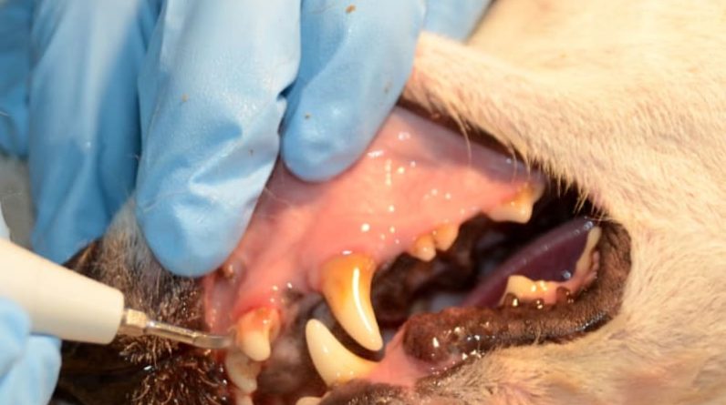
Photo of a pet skull with the left maxillary and mandibular permanent teeth numbered according to the modified Triadan system. Canines have 42 permanent teeth (12 incisors, 4 canines, 16 premolars, and 10 molars). The incisors (101– 103, 201– 203, 301– 303, 401– 403) and canine teeth (104, 204, 304, 404) are single rooted. In the maxillary arch, the very first premolars (105, 205) have 1 root, the second and 3rd premolars (106, 107, 206, 207) have 2 roots, and the fourth premolars (108, 208) and very first and second molars (109, 110, 209, 210) have 3 roots. In the mandibular arch, the first premolars (305, 405) have 1 root; the 2nd, 3rd, and fourth premolars (306– 308, 406– 408) and the very first and 2nd molars (309, 310, 409, 410) have 2 roots; and the third molars (311, 411) have 1 root … read more
Thanks to Dr. Maria Soltero-Rivera
Dentition, horse
Schematic of the dentition of a horse (Equus caballus). Illustrated are the anatomical functions of particular teeth, as well as the variations observed on the occlusal surfaces of the various kinds of teeth and as a function of age … learn more
Illustration by Dr. Gheorghe Constantinescu.Dentition, donkey Photographs of a donkey
skull with the maxillary and mandibular irreversible teeth numbered according to the modified Triadan system … learn more Courtesy of the Archaeozoological Reference Collection of the Institute of Topographic Anatomy, University of Veterinary Medication, Vienna.Dentition, small ruminants Schematic of the dentition of a small
ruminant. Illustrated are the
structural features of specific teeth, in addition to the variations observed on the occlusal surface areas of the different types of teeth and as a function of age … learn more Illustration by Dr. Gheorghe Constantinescu.Dentition, pig Schematic
of the dentition
of a pig( Sus scrofa domesticus), consisting of the anatomic features of specific teeth. Illustration by Dr. Gheorghe Constantinescu.For More Info Discover the veterinary topic of Dentition and Dental Classification of Animals.
Discover particular details on this topic and
related subjects from the
Merck Veterinarian Manual.